Cancer is a deadly disease that affects millions of people around the world, and early detection is vital. Medical technologies used in the diagnosis and treatment of cancer are constantly evolving and scintigraphy in the field of nuclear medicine is an important part of these technologies. Scintigraphy is a sensitive and reliable imaging technique that helps detect cancerous cells and determine the spread of the disease. In this article, we will try to give you information about what it is, what it does, and how it is performed.
- What Is Scintigraphy?
- What Does Scintigraphy Do?
- What Is Scintigraphy Used For?
- How Is Scintigraphy Performed?
- Is Scintigraphy Safe?
- Benefits of Scintigraphy
- What Are The Risks of Scintigraphy?
- How Is Scintigraphy Performed?
- Nuclear Scintigraphy
- How Does Nuclear Scintigraphy Work?
- What Are The Types of Scintigraphy?
- When Are the Scintigraphy Results Available?
- What Are The Side Effects of Scintigraphy?
- What To Do Before Scintigraphy?
- Why Is Scintigraphy Needed?
- Which Diseases Is Scintigraphy Used For?
- Types Of Cancer For Which Bone Scintigraphy Is Most Commonly Used
- How Is Bone Scintigraphy Performed?
- Scintigraphy Treatment Prices in Turkey
What Is Scintigraphy?
Scintigraphy is an imaging method used in the field of nuclear medicine in which radioactive substances are injected into the body and the functions of organs and tissues are examined. In this method, the radioactive substance is selected according to the diseased area and after being injected into the body, images are obtained with a gamma camera. This is a method for examining the internal structure of the body and the functions of organs and is particularly effective in detecting cancer cells and determining metastasis status.
What Does Scintigraphy Do?
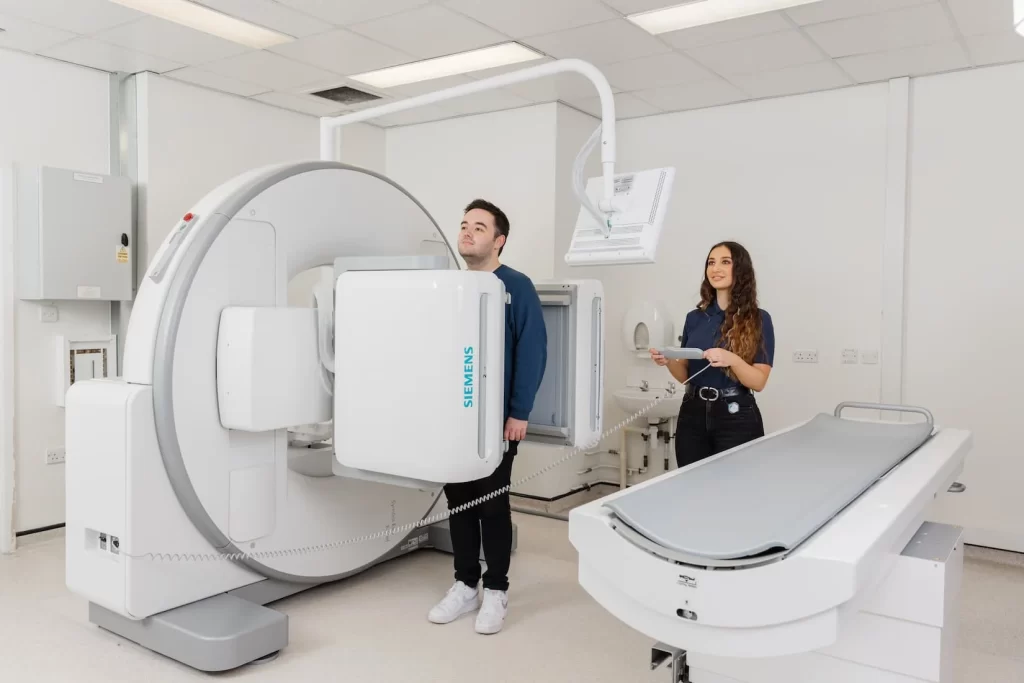
This plays an important role in the diagnosis and treatment of cancer. Detecting cancer cells helps to diagnose the disease at an early stage and determine appropriate treatment methods. It is also effective in determining the spread of the disease (metastasis). This examines the functions of many organs such as the thyroid, kidney, bone, heart, and brain, allowing the detection of cancerous cells occurring in these organs.
What Is Scintigraphy Used For?
This is a medical imaging technique that uses radioactive materials to create images of the body’s internal organs and tissues. The radioactive materials, called radiopharmaceuticals, are injected into the bloodstream or swallowed. The radiopharmaceuticals then accumulate in the organs or tissues of interest, where they emit gamma rays. These gamma rays are detected by a special camera, which creates images of the organs or tissues.
Scintigraphy is used to diagnose a variety of conditions, including:
- Cancer
- Heart disease
- Stroke
- Infections
- Thyroid problems
- Bone disorders
- Kidney disease
- Liver disease
- Gastrointestinal disorders
Scintigraphy can also be used to:
- Assess the function of an organ or tissue
- Monitor the progression of a disease
- Evaluate the response to treatment
- Guide surgery or other procedures
How Is Scintigraphy Performed?
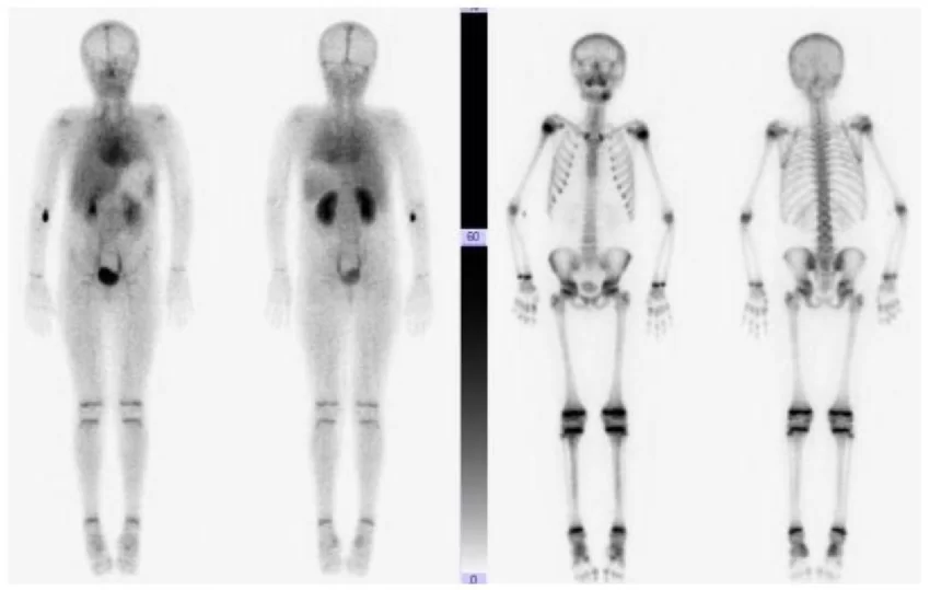
This is a relatively painless procedure. The patient will be given a radiopharmaceutical, either by injection or by mouth. The radiopharmaceutical will then be absorbed into the bloodstream or tissues. The patient will then lie down on a table under a special camera. The camera will take pictures of the body as the radiopharmaceutical moves through it. The procedure usually takes about 30 minutes.
Is Scintigraphy Safe?
Scintigraphy is generally considered to be a safe procedure. However, there is a small risk of side effects from the radiopharmaceuticals. These side effects can be mild, such as nausea or vomiting, or more serious, such as allergic reactions. The risks of this are usually outweighed by the benefits of the procedure.
Benefits of Scintigraphy
Scintigraphy is a medical imaging technique that uses radioactive materials to create images of the body’s internal organs and tissues. The radioactive materials, called radiopharmaceuticals, are injected into the bloodstream or swallowed.
The radiopharmaceuticals then accumulate in the organs or tissues of interest, where they emit gamma rays. These gamma rays are detected by a special camera, which creates images of the organs or tissues.
Scintigraphy has many benefits over other imaging techniques. Here are some of the most important benefits:
- Highly sensitive: It is a very sensitive technique that can often diagnose diseases that are not visible with other imaging methods. This is because the radiopharmaceuticals used in this are attracted to specific tissues or organs, making them easier to see on the images.
- Non-invasive: It is a non-invasive procedure, which means that it does not require any surgery or other invasive procedures. This makes it a safe and comfortable option for patients.
- Quick and easy: It is a relatively quick and easy procedure. The patient will typically be injected with the radiopharmaceutical and then lie down on a table under a special camera for about 30 minutes.
- Affordable: It is a relatively affordable imaging technique. This makes it a good option for patients who are looking for a cost-effective way to diagnose or monitor a disease.
What Are The Risks of Scintigraphy?
Scintigraphy is a generally safe procedure, but there are some risks associated with it.
These risks include:
- Radiation Exposure: The radiopharmaceuticals used in scintigraphy emit radiation. This radiation can pose a small risk of cancer, especially if the procedure is performed multiple times.
- Allergic Reaction: The radiopharmaceuticals used in it can cause an allergic reaction in some people. This reaction can be mild or severe.
- Other Side Effects: Other side effects of scintigraphy are rare, but they can include nausea, vomiting, headache, and dizziness.
The risks of scintigraphy are usually outweighed by the benefits of the procedure. However, it is important to talk to your doctor about the risks and benefits before you have the procedure.
Here are some additional things to keep in mind about the risks of scintigraphy:
- The amount of radiation exposure from it is usually very low. However, the risk of cancer increases with the amount of radiation exposure.
- The risk of an allergic reaction to the radiopharmaceuticals is also very low. However, it is important to tell your doctor if you have any allergies before you have the procedure.
- Other side effects of it are rare. However, it is important to be aware of them so that you can seek medical attention if they occur.
- If you are considering it, it is important to talk to your doctor about the risks and benefits of the procedure. Your doctor can help you decide if it is the right test for you.
How Is Scintigraphy Performed?
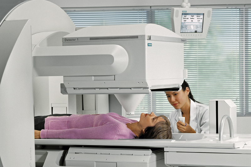
Scintigraphy begins with the injection of a radioactive substance into the body. This substance is usually injected intravenously and is selected according to the diseased area in the body. After the radioactive material is injected into the body, the patient may need to wait for a while. This is necessary for the substance to be distributed in the body and reach the target organ.
After the waiting period is over, the patient is placed in front of a device called a gamma camera. The gamma camera detects the rays emitted by the radioactive substance and obtains images. These images are evaluated by the doctor to get information about the condition of the disease and the level of spread. It is important that the patient does not feel discomfort or move during scintigraphy, as movement can reduce image quality and make it difficult to obtain accurate results.
Nuclear Scintigraphy
Nuclear scintigraphy is a medical imaging technique that uses radioactive materials to create images of the body’s internal organs and tissues. The radioactive materials, called radiopharmaceuticals, are injected into the bloodstream or swallowed. The radiopharmaceuticals then accumulate in the organs or tissues of interest, where they emit gamma rays. These gamma rays are detected by a special camera, which creates images of the organs or tissues.
How Does Nuclear Scintigraphy Work?
Nuclear scintigraphy is a relatively painless procedure. The patient will be given a radiopharmaceutical, either by injection or by mouth. The radiopharmaceutical will then be absorbed into the bloodstream or tissues. The patient will then lie down on a table under a special camera. The camera will take pictures of the body as the radiopharmaceutical moves through it. The procedure usually takes about 30 minutes.
The radiopharmaceuticals used in nuclear scintigraphy are very small and do not cause any harm to the body. They are attached to molecules that are specific to certain organs or tissues. This allows the radiopharmaceuticals to accumulate in the desired area, making it easier to see on the images.
The images produced by nuclear scintigraphy are called scintigrams. Scintigrams are often used in conjunction with other imaging methods, such as X-rays, CT scans, and MRI scans, to provide a more complete picture of the patient’s condition.
What Are The Types of Scintigraphy?
Scintigraphy is an imaging method that uses radioactive substances and shows the functions of organs in the body. It is used to evaluate the functioning and structure of organs in the body. This method provides important information for the diagnosis and treatment of organ diseases. Scintigraphy types are as follows;
Bone Scintigraphy: It is used to detect bone damage, fractures, infections, and tumors. It also has an important place in the diagnosis and follow-up of bone metastases.
Thyroid Scintigraphy: It is used to evaluate the functions and structures of the thyroid gland. Nodules, inflammation, and cancerous cells in the thyroid gland can be detected with this method.
Parathyroid Scintigraphy: It is used to evaluate the functions and structures of the parathyroid glands. Adenomas, hyperplasia, and cancerous cells in the parathyroid glands can be detected with this method.
Myocardial Perfusion Scintigraphy: It is used to evaluate the blood supply and function of the heart muscles. It is used in the diagnosis and follow-up of heart diseases such as coronary artery disease, heart attack, and heart failure.
Renal Scintigraphy: Used to evaluate the function and structure of the kidneys. It is used to diagnose and monitor kidney diseases such as kidney failure, kidney stones, and kidney infections.
Lung Scintigraphy: Used to assess the function and structure of the lungs. It is used in the diagnosis and follow-up of lung diseases such as pulmonary embolism, lung cancer, and lung infections.
When Are the Scintigraphy Results Available?
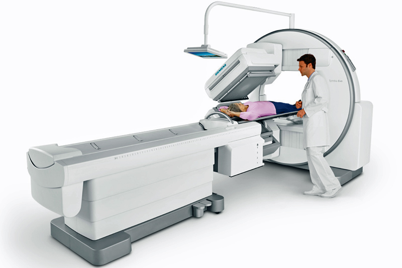
Scintigraphy is performed by waiting for a certain period of time after the radioactive material is injected into the body. This time may vary depending on the radioactive material used and the organ to be examined. The procedure time usually varies between 30 minutes and 2 hours. These results are evaluated by a nuclear medicine specialist after the procedure is completed. The results are usually available and reported on the same day. However, in some cases, it may take several days for the results to be evaluated and reported.
What Are The Side Effects of Scintigraphy?
Scintigraphy is generally safe and does not cause serious side effects. However, some people may experience allergic reactions to the radioactive material. This is very rare and usually has mild symptoms. There may also be temporary pain or redness in the area where the radioactive material is injected.
In terms of radiation exposure, scintigraphy uses low doses of radiation and therefore the risk is minimal. However, pregnant and breastfeeding women are advised to avoid scintigraphy. It is also important to drink plenty of water and urinate to flush the radioactive material out of the body. In this way, radiation exposure can be further reduced.
What To Do Before Scintigraphy?
Scintigraphy is a medical imaging method that uses radioactive substances called radionuclides and evaluates the functions of organs in the body. Patients need to make some preliminary preparations before the scintigraphy method. The things to be considered before the scintigraphy method can be explained as follows;
- You can continue or stop using your medications with the knowledge of your doctor before it. Inform your doctor about all medications, vitamins, and supplements you are taking.
- A fasting period of 4-6 hours is usually required before the scintigraphy method. You can only drink water during this time. If your doctor recommends a special diet, you should follow it.
- Since scintigraphy uses radioactive substances, it is important that pregnant or breastfeeding women inform their doctor. If necessary, the doctor may recommend a different imaging method.
- Tell your doctor if you are allergic to radioactive substances or medicines. In this case, the doctor will take the necessary precautions before it.
It is recommended that you wear comfortable clothes and, if possible, no metal accessories during it. Metal accessories can interfere with the imaging process.
Why Is Scintigraphy Needed?
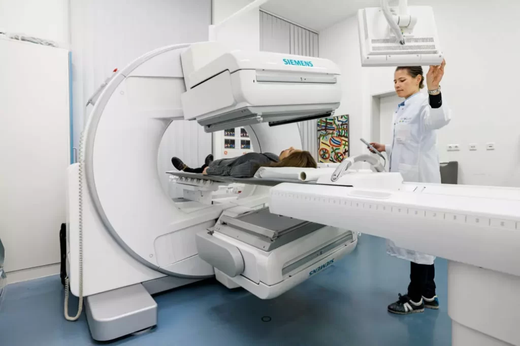
Scintigraphy is used to evaluate the function of organs in the body and to help diagnose many diseases. The situations where the Scintigraphy method is needed are as follows;
- It is used to examine the function of organs such as the kidney, thyroid, liver, and heart.
- It is used to diagnose and control the spread of cancers, especially thyroid, breast, prostate, and lung cancer.
- It is used to detect bone fractures, infections, tumors, and inflamed areas.
- It helps to identify areas of inflammation and infection in the body.
Which Diseases Is Scintigraphy Used For?
The scintigraphy method is used in the diagnosis and treatment of many diseases. Here are the diseases that can be diagnosed with it;
- It is used to evaluate the functions of the thyroid gland and to diagnose thyroid diseases such as thyroid nodules, goiter, and Graves’ disease.
- It is used to diagnose bone diseases such as bone fractures, bone tumors, osteoporosis, and Paget’s disease.
- It is used to diagnose and control the spread of cancers, especially thyroid, breast, prostate, and lung cancer.
- It is used to diagnose heart diseases such as heart muscle damage, coronary artery disease, and heart failure.
- It is used to diagnose kidney diseases such as kidney stones, kidney failure, and hydronephrosis.
- It is used to diagnose liver and spleen diseases.
- It is used to diagnose brain diseases such as Parkinson’s disease and Alzheimer’s disease.
Types Of Cancer For Which Bone Scintigraphy Is Most Commonly Used
Bone scintigraphy is an imaging method that shows abnormal changes occurring in the bones. This test becomes particularly important if cancer cells have spread to the bones. Bone scintigraphy can be used for the early detection of metastases (the spread of cancer from another organ to the bones).
The most common types of cancer for which bone scintigraphy is used are as follows;
- Prostate cancer is one of the most common types of cancer in men. In advanced cases of prostate cancer, cancer cells can spread to the bones. Therefore, bone scintigraphy plays an important role in the staging and treatment planning of prostate cancer.
- Breast cancer is the most common type of cancer in women. In advanced breast cancer, cancer cells tend to spread to the bones. Bone scintigraphy is used in the staging and treatment of breast cancer.
- Lung cancer is one of the deadliest cancers worldwide. Cancer cells can spread from the lungs to the bones. In this case, bone scintigraphy is used in the follow-up and treatment of lung cancer patients.
- Thyroid cancer originates from the thyroid gland, which is primarily located in the neck region. In advanced cases of thyroid cancer, cancer cells can spread to the bones. Bone scintigraphy is an important tool in thyroid cancer staging and treatment planning.
- Multiple myeloma is a type of blood cancer caused by the abnormal proliferation of plasma cells. In patients with multiple myeloma, abnormal changes in the bones and fractures can occur. Therefore, bone scintigraphy is used in the diagnosis and treatment of multiple myeloma patients.
How Is Bone Scintigraphy Performed?
Bone scintigraphy is a medical test that obtains an image of the bones in the body using a radioactive substance. The bone scintigraphy procedure can be explained step by step as follows;
Before the procedure begins, the patient is injected with a radioactive substance. This substance spreads to the bones in the body, allowing images showing abnormal changes to be obtained.
A period of time is allowed for the radioactive substance to spread to the bones in the body. This time usually varies between 2 and 4 hours.
After the radioactive material has spread to the bones, a special camera is placed on the patient. This camera detects the rays emitted by the radioactive material and creates images of the bones.
The images are evaluated by a radiologist. By detecting abnormal changes in the images, the radiologist determines whether cancer cells have spread to the bones.
Based on the results of the bone scintigraphy, the doctor creates a treatment plan for the patient. If the cancer cells have spread to the bones, the patient may receive chemotherapy, radiotherapy, or other treatment methods.
Scintigraphy Treatment Prices in Turkey
Turkey has managed to announce its name to the world with its investments and studies in the field of health. Especially the latest technological devices used in diagnosis and treatment procedures have been a beacon of hope for many diseases. However, there has been an increase in health tourism in Türkiye.
- Hospitals are large, clean, spacious, and fully equipped in terms of technological equipment.
- Turkish doctors are specialized, successful, and skilled in their fields.
- Nurses and carers are friendly and compassionate.
- Finding answers to the questions asked quickly and accurately.
- Patience and understanding of all staff, including the intermediary company dealing with the patient.
- Turkey offers holiday opportunities with its natural and historical beauties.
- Easy transportation.
- Diagnosis, treatment, accommodation, eating, drinking, dressing, and holiday needs can be met at affordable prices.
Such situations are shown among the reasons for preference. Scintigraphy Treatment Prices in Turkey
We can see that patients and relatives of patients who want to come to Turkey are doing research. However, it would not be right to give clear price information at this stage. Many factors such as the type of disease, stage, diagnosis process, treatment process, and stay in Türkiye affect the price issue. If you want to get more detailed price information, you can contact us. In addition, if you come to Turkey for treatment through us, we can facilitate your visa application process with the invitation letter sent by us to the consulate.

Vimfay International Health Services