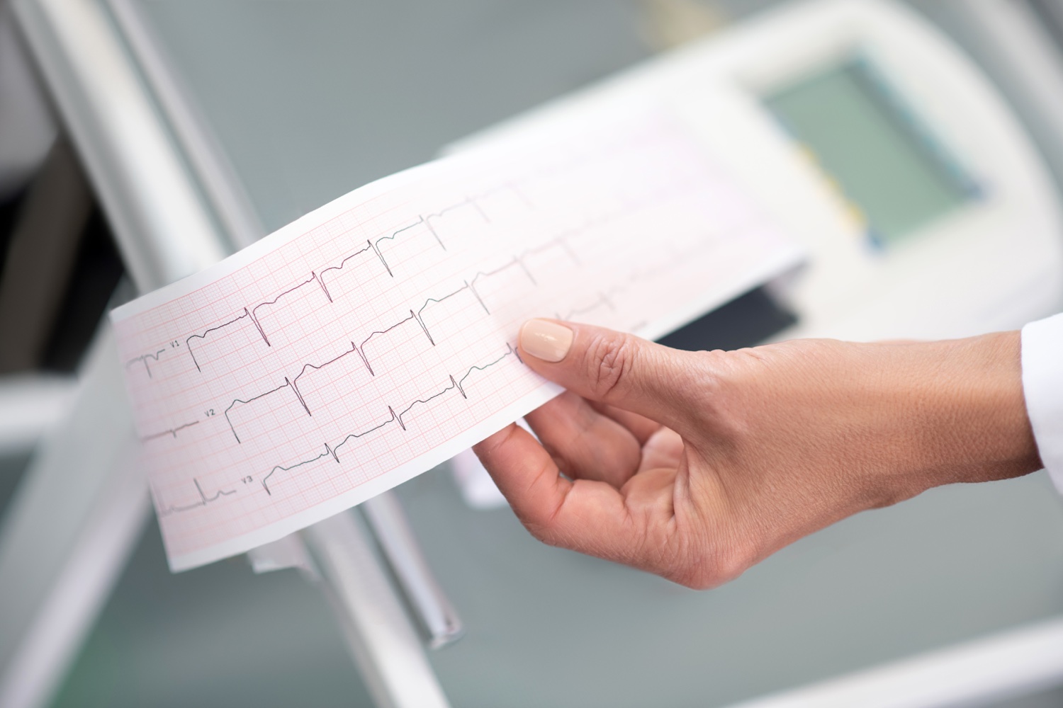
Cardiology is the branch of medicine that studies diseases related to the heart and circulatory system. The heart pumps the clean blood needed by each unit to function. At the same time, the heart must send the blood used and polluted in the body to the lungs to be cleaned. Symptoms such as chest pain, palpitations, dyspnea (shortness of breath), syncope (fainting), and nocturnal urination are often seen in individuals with heart disease. Current methods used in the diagnosis of heart diseases can be listed as electrocardiography (ECG), echocardiography (ECHO), chest X-ray, exercise test, coronary angiography, and cardiac catheterization. The most common heart diseases are hypertension, heart failure, and congenital heart anomalies.
Hypertension

Tension is defined as the blood pressure applied to the vessel walls. Hypertension (high blood pressure) is the condition in which the blood pressure applied to the walls of the arteries carrying blood from the heart to the whole body is above normal limits. The two most important factors affecting the blood pressure value of the person are the lumen width of the arteries and the amount of blood pumped by the heart. As the amount of blood pumped by the heart increases and the lumen width of the arteries decreases (the arteries narrower), blood pressure rises. The blood pressure value measured when the heart contracts to pump blood is called systolic pressure, while the blood pressure value measured when it relaxes to allow blood to fill inside is called diastolic pressure. The normal systolic blood pressure value for healthy adults is 120 mmHg (millimeters of mercury), and the diastolic blood pressure value is 80 mmHg. Values measured above 120/80 mmHg are determined as pre-hypertension.
Individuals with hypertension can continue their lives without symptoms for a long time. However, even asymptomatic cases of hypertension can cause deterioration in the vessel walls and heart muscle. Hypertension may develop spontaneously without any reason (primary hypertension), or it may develop due to kidney disease, thyroid problems, obstructive sleep apnea, or medications used by the individual (secondary hypertension). Determining risk factors for hypertension can be listed as age, overweight, sedentary lifestyle (inactivity), a diet rich in salt, smoking, alcohol use, and high stress. Some of the complications that may occur due to hypertension are atherosclerosis, metabolic syndrome, vision loss, kidney diseases, and cognitive disorders.
For the diagnosis of hypertension, the blood pressure of the individual is evaluated by measuring at least three different times. In addition, in some cases, it may be requested to follow up with blood pressure measurement at home. The diagnosis is confirmed by blood and urine tests and echocardiogram evaluation.
Regular use of prescribed drugs is sufficient for the treatment of hypertension. The first preferred drug group is calcium channel blockers. Beta-blockers may be preferred in cases of hypertension that cannot be controlled with the use of calcium channel blockers. Additional diagnostic methods are applied for hypertension cases that cannot be controlled by regular use of prescribed drugs. Cases of hypertension that cannot be controlled despite three different drugs used (at least one of the drugs must be in the diuretic drug group) or that can be controlled with four drugs used simultaneously are called resistant hypertension.
There are some elements that individuals with hypertension should pay attention to throughout their life. Regular physical activity habits, following heart-friendly diets (including low-fat fish and poultry), maintaining a healthy weight, not smoking, and developing ways to cope with stress are some of them.
Heart Failure

Heart failure is defined as the inability of the heart to meet the amount of blood needed by the body and the inability to pump blood to the body. Heart failure, which is a progressive and chronic disease, can develop due to different reasons. These reasons are diseases such as previous myocardial infarction (heart attack), heart valve diseases, and hypertension.
Patients with heart failure most frequently apply to clinics with complaints of dyspnea (shortness of breath), prolonged edema, and fatigue. Patients’ complaints of dyspnea often develop during movement, and some patients may also develop orthopnea, which is a very difficult situation to cope with. Orthopnea is defined as shortness of breath when lying on the back. Edema complaints of the patients are seen around the ankles, especially in the lower extremities.
Medical treatment is preferred primarily in the treatment of heart failure. Angiotensin receptor blockers (ARB), angiotensin-converting enzyme inhibitors (ACEI), diuretics, and beta-blockers are used in medical treatment. ARB group drugs are drugs that act by lowering blood pressure. ACEI group drugs have a strengthening effect on the heart muscle. Diuretic drugs affect the removal of the accumulated fluid from the kidneys by filtration in patients suffering from edema. Beta-blockers, on the other hand, slow down the heart rate and have a vasodilator effect. Thus, they provide better nutrition to the heart muscle.
In cases where medical treatment is insufficient in the treatment of heart failure, surgical treatment can be performed. These surgeries and assistive devices are coronary artery bypass surgery, heart valve replacement, heart valve repair, ICD (implantable cardioverter-defibrillator), dual-chamber pacemaker, and heart transplant surgeries.
Coronary artery bypass surgery is a surgery applied to the arteries that feed the heart itself (coronary arteries). The artery with narrowing or occlusion from these arteries is bypassed and the heart is fed by another artery. Thus, the number of nutrients and oxygen going to the heart muscle is kept at an optimal level.
Heart valve surgeries are performed by repairing a dysfunctional heart valve or replacing it with prosthetic valves.
ICDs are special devices that have been developed to intervene in rhythm disorders in the heart and can provide shock therapy. These devices are usually placed under local anesthesia about 2 cm below the collarbone in the chest area of the person. The electrodes of the device are passed through a vein from the arm to the heart and delivered to the heart chambers.
Dual-chamber pacemaker is preferred in patients who have problems with the conduction system of the heart. With the electrodes placed in the right and left ventricles, harmonious contraction of the ventricles is ensured.
Heart transplant surgery is preferred in patients who cannot find a solution with any medical treatment or assistive device surgery.
Atrial Septal Defect

An atrial septal defect is a congenital disease defined as the presence of a hole in the septum wall that separates the right and left atrium of the heart. Under normal conditions, the blood in the two atria does not mix, but in the case of ASD, some of the clean blood in the left atrium may pass into the right atrium. As the volume of blood in the right atrium increases, the volume of blood pumped from there to the lungs also increases. Over time, the increased blood volume in the right heart causes the right atrium and right ventricle to enlarge. This growth can lead to the development of heart failure and pulmonary hypertension. Atrial septal defects are examined in four groups primum, secundum, sinus venosus, and coronary sinus defects.
The complaints of patients with atrial septal defects vary depending on the size and localization of the hole. The diagnosis of ASD is made with the help of echocardiography Increased right ventricular volume loading on echocardiography is an important finding for the diagnosis of ASD. In case of insufficient echocardiography, transesophageal echocardiography, magnetic resonance imaging, and computed tomography methods can also be used. After determining the size, number, localization of the hole or holes, and their distance from each other by evaluation methods, the method to be used for closing the holes are decided.
It is possible to close the secundum type defect, which is one of the atrial septal defect types, without surgery using a catheter. During this procedure, the heart does not need to be stopped, and since the chest is not opened, cosmetic problems such as large scars and permanent deformities are not seen. Under local anesthesia, the right atrium is reached by entering through the femoral vein from the inguinal region, and the ASD is closed. If the diameter of the hole is larger than 26 mm and there is more than one hole, if the distance between the holes is less than 5 mm, the closing process is performed using a single device. If the hole or holes do not comply with the closure conditions with a single device, the heart is reached by using both veins in the inguinal region.
ASD closure is a safe treatment method and has potential complications. Incorrect placement of the device, displacement of the device, development of cardiac tamponade, rhythm disorders, development of thromboembolism, migraine, and transient ischemic attack are among the reported complications.
After the ASD closure procedure, the patients are followed up regularly. Patients are generally evaluated with echocardiography every six months for two years after the procedure. In addition, patients use aspirin for six months after the procedure.